HomeDiagnoseTherapySurgeryPhotosLinksPhysician
anatomyphotosvideoaudiocase histories

Case histories: Capillary ectasias


History

Complaints

  • 4th decade
  • female
  • elementary school music teacher
  • Two years of loss of singing capabilities for five months after school starts
  • definate improvement over the summer
  • Possible loss of high soft singing for a longer period
  • voice fatigue
  • previous ENT exam - told she had nodules - Rx Speech therapy - no help according to patient

Medical history

  • Soprano
  • 10 years vocal training
  • tobacco - none
  • alcohol - none
  • caffeine - none
  • reflux symptoms - mild
  • Medications - muscle relaxants

Character

  • talkativeness scale: 4
  • loudness scale: 4
  • vocal commitments
    • 6 hours talking at work
    • choir practice 3 hours per week
    • choir perfomance 1 hour per week

Vocal capabilities

  • Reading voice
    • clear sounding
  • Pitch range
    • Anchor pitch (F0) - F3#
    • Low pitch - F3#
    • High pitch - A5
    • pianissimo mucosal ceiling (onset delays and breathiness) - C5 onset delay, F5 loss of entrainment
    • diplophonia - no

Stroboscopic Examination of the Larynx

  • broad based swelling along both vibratory margins
  • hemorrhage on left noted during open phase of strobe
  • ectasia on right
  • clear and translucent
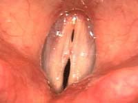
polyp and ectasia Stroboscopic view at F3#. This image was chosen at this low pitch on stroboscopic exam to highlight the capillaries within the vocal fold polyps. The ectatic vessels are visible centrally on the right in the polyp. On the left, the ectasia is visible in the posterior half of the polyp.
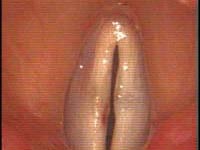
polyp and ectasia post speech therapy Post speech therapy - Stroboscopic view at F4#. There are typically two lips visible on stroboscopic exam. When the upper lip is at its most lateral, the medial surface of the vocal folds is sometimes visible as in this case. The ectatic vessels are visible as a pinpoint red area on the right in the larger polyp. On the left two ectasias have hemorrhaged.
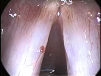
surgical view - polyp and ectasia Surgical view. Taken through a 3.5 mm - 30 degree endoscope. This scope allows viewing of the actual medial surface of the vocal folds. What was a somewhat vague spot on the above stroboscopic exam is clearly a well defined tortuous capillary.
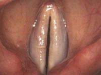
2 weeks post removal of polyp and ectasia Postoperative view. Two weeks after surgery. The patient is resuming singing.

Treatment

Medical

No medications were recommended.

Behavioral

Physician treatment information
Patient treatment information

Behavioral modification is the initial treatment of these mucosal lesions and is likely a lifelong treatment for this patient. It is seldom someone can change their personality but it is possible for someone to manage their behavior. She is a severely impaired singer, yet it is usually not the singing that is the problem. More often it is the amount of talking that goes on daily. She rates herself a 4 and 4 on the talkativeness and loudness scales, but does talk and sing extensively at school. This usage carries over into home life with choral practice and performance.

Many times managing talkativeness will reduce a vocal fold swelling to an acceptable size such that the voice becomes dependable and acceptable to the patient, though the capillary ectasia on the vibratory margin makes this a difficult managment problem since that will likely swell with moderate usage or rupture intermittantly.

Speech therapy was again tried and was very helpful in controlling the amount and style of vocal use yet failed to prevent repeated hemorrhage.

Surgical

Physician treatment information

After behavioral management surgery was indicated since the patient desired further improvement. Surgery is directed at removing only the mucosal lesion and preserving as much of the intermediate and deep layers of the vocal fold as possible.

With an appropriately delicate touch, these polyps were removed most successfully. Lasering of the vessel on the vocal fold margin successfully removed these ectasias. She experienced a substantial improvement.

Contact the author: James P. Thomas, MD

Updated 14 April 2004