Review of High-definition laryngoscopes Olympus vs. Pentax
Pentax vs. Olympus flexible chip HD laryngoscopes
I had a chance for one week (June 2012) to compare the Pentax high definition flexible chip laryngoscope - VNL-1590STi (which I have used for one year) with the Olympus high-definition flexible chip laryngoscope - ENF-VH, which came out in late 2011. The Pentax scope has been available for a longer period of time. My overall impression is at the bottom of this review.
My Voice Lab Recording setup
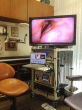
First a few details about my setup since I do not use the proprietary video capture system of either Pentax or Olympus. All of my equipment is set up on a tower with a flatscreen monitor on top. My MacBook Pro laptop sits in front of the stroboscope. Audio equipment is hidden in the back. tThe video processor is on the lower shelf.
Video capture: I capture endoscopic video images on my laptop computer (MacBook Pro 13inch - OS X 10.6.8) using Final Cut Pro 7.0.3. Consequently I need to ingest video through the FireWire port (IEEE 1394 interface). My video files are captured as .mov files which are easy to view, store or convert to other video formats. The files can easily be viewed on the desktop, in Quicktime, in iMovie, in Final Cut Pro or other video applications.
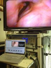
Video storage and backup: For those of you that worry about the transient nature of digital files, at the end of each patient exam I copy the captured video files from my laptop to a LaCie hard drive (10TB LaCie 4big Quadra) configured as RAID 10 storage - which saves two copies of each file on two drives simultaneously. Over the past 10 years, I have performed approximately 10,000 examinations of roughly 4 minutes of video each. The majority of them have been recorded in SD and I am able to store all of the files on two hard drives (~18 TB total). These two drives physically take up 220 x 196 x 173 mm (HxLxW) of space each. Approximately once a month, I make a backup onto another hard drive and physically store that drive off-site. I utilize CrashPlan to continuously back up daily the Final Cut Pro datafiles.
Level of definition: HD (high-definition) video produces significantly larger files than SD. How much more depends on how high the definition is and what compression algorithm is chosen. There is not yet a single standard for the term “high-definition video” or “HD video”. Two common HD standards are 720p and 1080i.
The Pentax EPKi video processor does conveniently have a DV output which can be plugged directly into an Apple laptop for capturing SD (standard definition) video. However, neither the Pentax nor the Olympus processors output high definition video through FireWire, which is Apples preferred method of ingestion of video. Since I cannot plug my laptop directly into either of the video processors for capturing an HD signal, I need some type of hardware conversion of the video signal and Olympus and Pentax have chosen different output methods.
Equipment compared:
I compared the Pentax VNL-1590 STi (HD ~720p) and the Pentax VNL-1170K (SD) to the Olympus ENF-VH (HD 1080i) and the ENF-V3 (square partial frame of HD 1080i). I also have a rigid 90° laryngeal endoscope which can be connected with a C-Mount to an HD camera. In order to capture HD, the most current video processor from each company is necessary. These processors also support their earlier SD video endoscopes.
Video processors:
The Pentax VNL-1590 STi high definition endoscope ($17,500 retail 2011) requires the Pentax EPKi video processor ($36,750 retail 2011). (Total retail cost: $54,250) The xenon light in the video processor are integrated into one unit. For the Pentax EPKi video processor, the only option is to capture the high definition signal from the DVI output. The other video outputs on the rear of the device are all SD or analog (S-video or Component-video).
The Pentax EPKi video processor outputs a computer graphics signal which is 1280x1024 pixels otherwise known as SXGA. I have not been able to find out what the actual resolution is of the chip on the endoscope. In order for me to capture this signal on my Macintosh, I need to convert it to a video format and 720p, which is 1280 x 720 pixels and 59.94 frames per second is the closest approximation to SXGA although I lose some of the vertical lines of resolution. I could upscale it to 1080i which is 1920 x 1080 pixels which generally yields a significantly larger file size depending on compression. In testing this out, I did not find a significant image difference, so I have elected to capture at 720p. The height to width ratio of HD video is 16:9 as compared to a ratio of 4:3 for SD video. My HD videos appear to have a slightly squished vertical appearance as recorded. This can be corrected afterwards in Final Cut Pro or when capturing or exporting still photos in Photoshop.
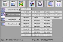
I use a Gefen DVI to HD-SDI Scaler ((SKU: EXT-DVI-2-HDSDISSL - $879 in 2011 at B&H Photo ) seen in the photo above sitting on top of that Pentax video processor. This device automatically detects the output from the DVI source. I then set the device to convert the computer graphics signal to a video signal and I have chosen 720p although 1080i and other formats are also available. Video from the Pentax EPKi via the Gefen Scaler is routed into an HD-SDI cable. in the photo below, in the lower left corner is the auto detected signal. In the lower right corner is my chosen output.
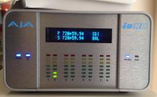
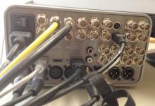
I then capture the HD SDI signal with an AJA Io HD ($3495 in 2011 at bhphotovideo.com) which integrates reasonably well with Final Cut Pro. This device auto detects the type of incoming video signal. The software with the AJA Io HD device is highly flexible with many (almost bewildering) choices for compression before output. I have settled on using the full resolution Apple ProRes 422 10 bit for 1080i capture, the Apple ProRes 422 for 720p and DV for SD video. Although all of the video could be captured at the highest resolution to avoid switching settings when changing endoscopes, file sizes might be unreasonably large for the lower resolutions. Consequently when I change in the scope, I change the video capture settings and Final Cut Pro.
Capturing the same test movie at the 3 resolutions for 1 minute consumed the following amounts of storage:
525i: 234 MB
720p: 624 MB
1080i: 748 MB
The Olympus high definition endoscope ENF-VH ($26,700 in 2012) is coupled to an Olympus OTV-S190 Visera Elite processor ($22,500 in 2012) which has a DVI output as well as a HD SDI (High-definition serial digital interface) output. Since the light is not integrated into the video processor as it is in Pentax, a separate light source is necessary. A Visera Elite Xenon light CLV-S190 was utilized ($12,500 in 2012). (Total retail cost: $61,700) There are also standard definition video outputs. For my use, the HD SDI output is more convenient for video capture saving me the need to send a signal through a scaler and I routed the Olympus video into the second HD SDI input on the AJA Io HD device. I then use the AJA software to switch between the two video processors or between the various resolutions that different endoscopes offer.
Video processor added features: I do not use any of the patient data features which allow you to type patient information into the video processor and have it displayed overlying the image. Since I show video images and print still photos from them, I prefer to keep all my video images anonymous and have the data stored as metadata. Then, should a video or photo presentation require patient privacy, the images of the larynx have no visual data present. I can use the Final Cut Pro program to overlay text later if desired.
My rigid 90° Stortz endoscope is connected to a Toshiba 9213HD Camera with a C-mount coupler (sold by Kay-Pentax in 2011 for $14825 retail) which records in 1080i HD. The output from this is routed through an HD-SDI cable into the AJA Io HD. We can compare the videos/photos obtained with each.
Video endoscopes:
The Pentax flexible chip laryngoscope (VNL-1590STi High-Definition Videonasolaryngoscope) is reported to have the following specifications:
Field of view: 100°
depth of field 3-50 mm
maximum diameter: 5.1 mm
tip diameter ?
working length 300 mm.
The Olympus high definition chip laryngoscope ENF-VH (Olympus calls it a Rhino-laryngo videoscope) is reported to have the following specifications:
Field of view: 110°
Depth of field: 3.5-50 mm
maximum diameter: 3.9 mm
tip diameter: 3.9 mm
working length: 300 mm
Using the equipment:
Size: The Pentax endoscope is 5.1 mm in diameter, which is large enough that it typically requires extra decongestant, topical anesthesia and patience during insertion through a patient's nose. Even then, the fit can be firm and in about 15% of adult patients I cannot insert the endoscope tolerably in the office. The Olympus endoscope at 3.9 mm in diameter slips easily into adults after my typical topical decongestion (3 squirts of phenylephrine with 4% lidocaine via a MAD device).
Additionally, when trying to move the endoscope in close proximity to the vocal cords without additional topical anesthesia on the larynx, the smaller Olympus HD scope can be maneuvered closer than the Pentax. Moving closer to the larynx effectively provides higher-resolution images of lesions on the vocal cord since more of the lesion fills more of the pixels of the endoscope improving the resolution of the pathology.
False color imaging: Both video endoscope processors allow quick color alteration of the video recordings through changing color filters with the click of a button on the endoscope. Pentax calls their false color imaging “iScan” and Olympus calls their false color imaging “Narrow Band Imaging” or NBI. Essentially a color filter is utilized to highlight the color red. The mucosa of the larynx is nearly translucent and blood vessels beneath surface are quite visible. Certain types of lesions will cause certain types of alterations in the blood vessels of the larynx. Recognizing and visualizing anatomic changes in the blood vessels allows the examiner to more easily visualize some pathology, particularly tumors.
Stroboscopy:
I use the current version of Kay-Pentax stroboscope (Model 9400 - retail $15,995 in 2011). Olympus provided an LED stroboscope (StrobeLED - CLL-S1) for comparison.
Olympus StrobeLED: The stroboscope utilizes a lapel microphone which picks up any sound in the room and flashes the strobe light. If only the patient is making sound, a very good image of the vocal fold edges oscillating is recorded. As soon as the patient stopped making sound, the light immediately stabilized maintaining a clear image.
I noticed some problems with the stroboscope. Even after white balancing the Olympus stroboscope, the images always had a yellow hue. This is not overly problematic and does not impede interpretation of the motion of the vocal cords. There is apparently a color mode adjustment for this which I learned about after the equipment was gone. The microphone is a lapel style mic and seem to pick up sound from the patient's voice very well.
I do have the sense that there is less light provided by the LED light and although the images appear to have the same light intensity, some of the brightness for the Olympus LED stroboscope comes from video gain. Consequently there is more video noise on the stroboscopic images obtained with the Olympus stroboscope relative to the Kay-Pentax stroboscope. The light for video recording with the Olympus stroboscope is slightly more stable than with the Kay-Pentax. I do not know the durability or longevity nor cost of replacement of the LED light source.
Kay-Pentax stroboscope: This stroboscope has been the gold standard in laryngology for the past 20 years. I have used it for the past 15 years. The xenon light is very bright and leads to clear images during the stroboscopy recording. A video sync cable is used between the video processor and the stroboscope.
It has a stethoscope as a sound source pickup. Placed on the patient’s neck, it is highly selective for picking up the sound produced by the patient’s vocal cords. External noise seldom interferes with this pickup, but placement on the neck can be sensitive and less than optimum placement sometimes leads to flickering of the image with black frames recorded on the video file. This will also tend to occur as the xenon light fades. My xenon light tends to last for approximately 18 months worth of examinations. It is important to note that it costs approximately $800 to purchase a new xenon light.
Sample case - vocal polyp:
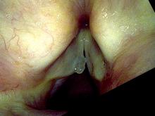
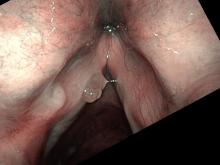
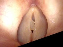
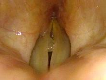
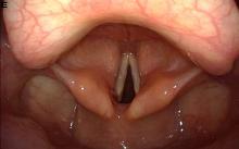
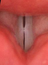
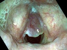
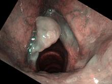
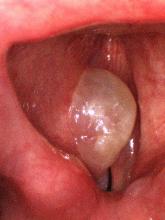
This patient has a unilateral, multilobulated polyp on the left vocal cord. The first image is viewed with the Pentax VNL-1590 STi at 720p
this image is viewed with the Olympus ENF-VH at 1080i
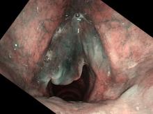
this image is viewed with the Olympus ENF-VH along with narrow band imaging at 1080i

This stroboscopy image is viewed with the Pentax VNL-1590 STi and the Pentax stroboscope at 720p
this image is viewed with the Olympus ENF-VH and the Olympus stroboscope at 1080i
vocal nodules:
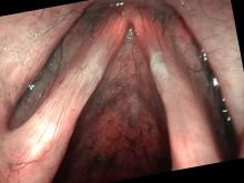
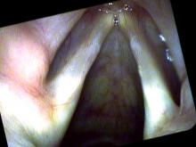
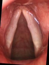
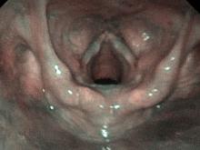
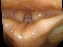
This youthful singer has bilateral vocal cord nodules. The 1st image is viewed with the Olympus HD endoscope at 1080i from above. In the 2nd image, the Olympus HD endoscope has been moved closer and the NBI color imaging has been engaged. The 3rd image is viewed with the Pentax VNL-1590 STi at 720p and the iScan color. The 4th image was obtained with the Olympus HD endoscope and the Olympus stroboscope. The 5th image was obtained with the Pentax HD endoscope and the Kay-Pentax stroboscope. The final 2 images were obtained with the rigid 90° endoscope and Toshiba processor during breathing and then on stroboscopy. The differences between the photos are more noticeable when viewed at their enlarged sizes.
Smoker's polyps:
This patient has bilateral smoker's polyps with the left being much larger than the right. The 1st image is viewed with the Pentax VNL-1590 STi at 720p and the iScan color. The next 2 images are viewed during breathing out and breathing in with the Olympus ENF-VH. The final image is viewed with the 90° rigid endoscope and Toshiba processor.
Olympus ENF-V3:
I was able to examine a 4-year-old child with vocal nodules with the Olympus ENF-V3, which is a smaller endoscope but still takes quite good images even though it only uses a portion of the high definition frame. The left photos taken with the NBI color feature engaged. The right photos taken with the Olympus stroboscope.
My impression:
Overall, my impression is that the Olympus ENF-VH provides an outstanding high-resolution image because of its 1080i output. Its 3.9 mm diameter allows for an easy and comfortable examination in almost any adult patient. Even without color filtering, the Olympus video processor tends to emphasize blood vessels more than the Pentax video processor. Until one gets used to the image, moving up from a fiber-optic endoscope to this HD video endoscope can erroneously suggest pathologic blood vessels in patients. However, when one is used to the type of image, the blood vessels are very important in identifying pathology. When the NBI is turned on, blood vessels turn quite black and distinct from the surrounding tissue. The resulting visualized vascular patterns of squamous cell carcinoma, laryngeal papilloma, dilated, ectatic or ruptured blood vessels within the vocal cord or within hemorrhagic polyps are very easily discerned. This is an extremely valuable addition to laryngeal imaging. Margins of tumors are very distinctly visualized.
This endoscope could probably typically be used in patients down to about 10 years old. If I had this endoscope, in my day-to-day workflow it would be my primary endoscope for laryngology. This does lead to significantly larger data files. Over the years, it does seem that storage technology has kept pace with imaging technology allowing for the progressively larger data files so I would not worry about this too much. I would rate it a 10 out of 10 for present-day (2012) high-definition technology. I would have to say that it is now the standard against which other laryngeal video endoscopes will be measured.
Caution: For several years Olympus has produced a high definition video processor without a compatible high definition flexible laryngoscope, but they marketed their standard definition laryngoscope with the high definition processor (which would enlarge the standard 720x480 image to the 1920x1080 size) using the term "high resolution". This has been confusing to buyers of the product. It remains a standard definition image stored as a larger file size.
The Olympus ENF-V3 endoscope at 2.6 mm in diameter was used in a four-year-old child for excellent quality images even though they only use a portion of the HD frame. With this as a secondary endoscope when the larger ENF-VH cannot be inserted, almost anyone who can sit still enough for an examination can have a clear, high-quality image of their larynx. It is an excellent second laryngoscope.
The Pentax VNL-1590 STi is an excellent quality HD endoscope for visualizing the larynx. The HD resolution is less than that of the Olympus ENF-VH. The 5.1 mm diameter is a significant factor limiting its use to adults and I am unable to place it through the nose in about 15% of adult patients. For me, it has become a secondary laryngoscope in my workflow. I typically look first with a standard definition flexible laryngoscope (Pentax VNL-1170K - 3.7 mm working diameter) which is easy to use and keeps my video data files small. Then for patients with pathology that warrants a closer look, I take the additional time to insert the larger Pentax HD laryngoscope. This is a somewhat slower workflow than I would have with the Olympus equipment. The lack of an HD SDI port on the compatible video processor requires the purchase of an additional video scaler adding to the cost. Having used a Pentax VNL-1170K endoscope for many years before requiring even minor service, I suspect that the durability of the Pentax HD endoscope will be similar, which is to say excellent. I have already used this HD endoscope daily for more than a year. The power supply failed and was replaced under warranty at about 14 months. I would rate it a 7/10 for present-day (2012) high-definition technology. Using each company's stated retail cost, the Pentax HD setup is about 10% less expensive than Olympus.

