HomeDiagnoseTherapySurgeryPhotosLinksPhysician
anatomyphotosvideoaudiocase histories
Case histories > PABRLN > EMG findings
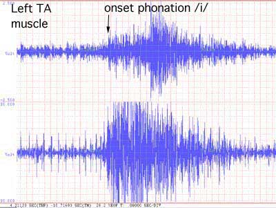
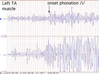
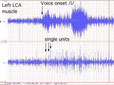
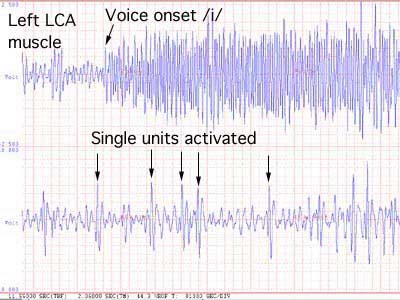
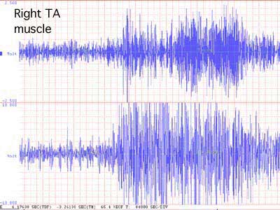
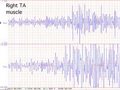
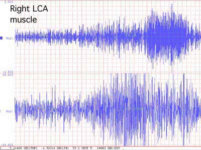
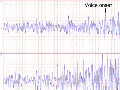
The following two channel recordings have the voice channel on the top row and the monopolor needle electrode recording on the second line. A Nicolet monopolar hollow needle electrode was inserted trans laryngeal through the cricothyroid membrane. Ground and reference electrodes were on the left side of the neck. The left frame is compressed 4 times the right frame to show a longer time segment. The right frame shows the individual waveforms clearer. Clicking on a diagram will produce a larger version. The EMG was recorded using a MA-100 Multichannel EMG by Motion Lab Systems. Data acquisition was through a Dateq DI-720 board on a Celeron 400 mHz PC.

LCA muscle EMG LCA close up view
The left TA muscle shows a good interference pattern.

LCA muscle EMG LCA close up view
Left LCA muscle position was confirmed by
  1. angling the needle lateral to the TA, yet just medial to the thyroid cartilage.
  2. checking high and low pitch for comparison with the CT muscle
    • the CT activated appropriately with a larger amplitude interference pattern with high pitched phonation
  3. verifying that the muscle activity in this case was different than the TA activity
  4. activity was compared to a similarly placed needle on the right side
  5. additional information could be obtained by doing a sniff /i/ maneuver to quiet all activity just prior to phonation

right TA EMG right TA close up view
Right TA muscle

right LCA EMG right LCA close up view
Right LCA muscle
Contact the author: James P. Thomas, MD
Updated 18 March 2001