vocal cord nodules recur after excision

Click on the image to see full-size version.

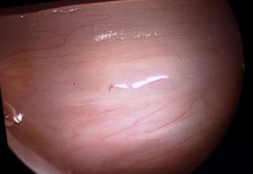
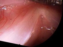
Vocal cord nodules during abduction, Vocal cord nodules during stroboscopy, Vocal cord nodules , 70° endoscopic surgical view of left vocal cord nodule, 70° endoscopic surgical view of left vocal cord nodule, 70° endoscopic surgical view of right vocal cord nodule, vocal cord nodules, microlaryngoscopy, microlaryngoscopy with excision of left vocal cord nodules with laser, 70° endoscopic surgical view of left vocal cord nodule after excision, 70° endoscopic surgical view of left vocal cord nodule after excision, 70° endoscopic surgical view of right vocal cord nodule after excision, 30° endoscopic surgical view of vocal cord nodules after excision, one week after microlaryngoscopy with excision of vocal cord nodules and voice rest, Three weeks after excision of vocal cord nodules, Three weeks after excision of vocal cord nodules, Two months after excision of vocal cord nodules, they are starting to recur, stroboscopy two months after excision of vocal cord nodules, 4 months after excision of vocal cord nodules, stroboscopy 4 months after excision of vocal cord nodules

Elizabeth is in her 20s and began singing about 3 years ago. About 1 year ago, she noted some roughness in her singing voice. She loves to talk and rates herself a 7/7 on the talkativeness scale and a 7/7 on the loudness scale. Here are her vocal cords when I met her.
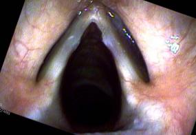
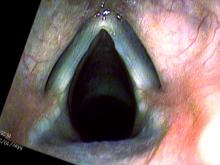
Vocal cord nodules during abductionVocal cord nodules during stroboscopy

On the left with the vocal cords open (abduction), there are fairly large white calluses in the center of each vocal cord with the right one being slightly larger. The vocal impairment they create is visible on the right. As she brings her vocal cords together at high pitch, vocal nodules touch each other splitting her vocal cord into 2 vibrating segments.

She went through with therapy and then subsequently microlaryngoscopy to remove the vocal cord nodules.

Surgery

Vocal cord nodules 70° endoscopic surgical view of left vocal cord nodule70° endoscopic surgical view of left vocal cord nodule70° endoscopic surgical view of right vocal cord nodulevocal cord nodules, microlaryngoscopymicrolaryngoscopy with excision of left vocal cord nodules with laser

The vocal cords are viewed with 30° and 70° endoscopes as well as a microscope. Then the nodules are excised by pulling the nodule away from the vocal cord and cutting with a CO2 laser.

70° endoscopic surgical view of left vocal cord nodule after excision70° endoscopic surgical view of left vocal cord nodule after excision70° endoscopic surgical view of right vocal cord nodule after excision30° endoscopic surgical view of vocal cord nodules after excision

At the conclusion of surgery, there is a depression where the mucosa or epithelium has been removed on each side.

She rested her voice completely for one week.

one week after microlaryngoscopy with excision of vocal cord nodules and voice rest

After 2 weeks of voice rest and one of speaking:

Three weeks after excision of vocal cord nodulesThree weeks after excision of vocal cord nodules

 vocal swellings are starting to reappear.

Two months after surgery, further voice therapy and rather active voice use:

Two months after excision of vocal cord nodules, they are starting to recurstroboscopy two months after excision of vocal cord nodules

Four months after surgery and continued voice use:
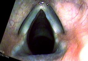
4 months after excision of vocal cord nodulesstroboscopy 4 months after excision of vocal cord nodules

She is now able to sing 7 semitones higher before her recurrent vocal swellings touch and impair her singing. One of the most important things about vocal overdoers is that the operation is on the vocal cords and not the patient's brain. It takes a great deal of vocal behavior modification to reduce vocal overuse, otherwise vocal swellings/nodules tend to recur.