Leukoplakia - verrucous keratosis
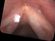
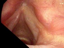
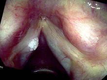
Leukoplakia of the left vocal cord. 40 years ago he quit smoking after 10 years of cigarettes. He came in with a lesion on his left vocal cord.
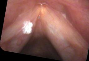
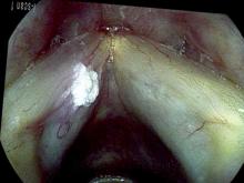
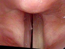
After one month, the lesion was still there, perhaps a bit larger. It is viewed here with false color imaging to highlight the blood vessels.
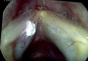
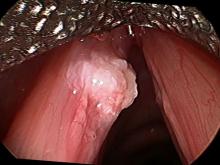
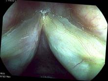
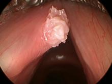
At surgery, a close-up of the lesion with a 30° endoscope looks like this.
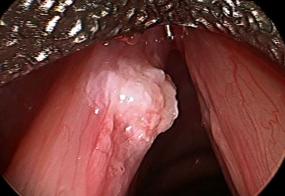
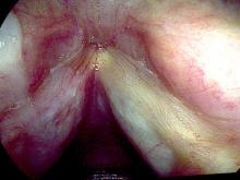
The pathology report described this as verrucus keratosis. One month after surgery there is some scar tissue present.
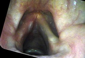
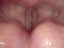
Two months after surgery there is still some white tissue present which could be either scar or residual lesion. It projects a little bit from the edge of his vocal cord and interferes with vibrations during stroboscopy.
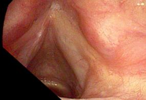
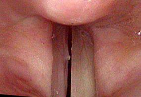
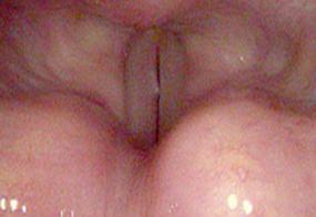
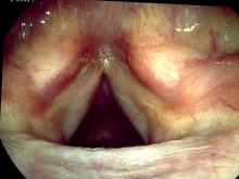
At his six month follow-up, it appears to be scar tissue.
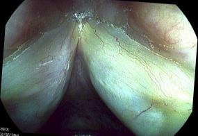
1 1/2 years later his voice was hoarse again. The white growth on his left vocal cord appeared quite similar to previous photos.
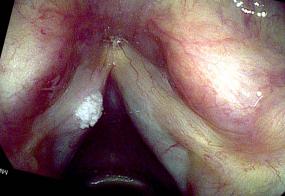
He had this removed again. At surgery, it appeared to be a white flaky, exophytic mass.
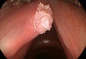
I incised deeper with this surgery. The pathology report again suggested that this was verrucous keratosis, a benign growth.
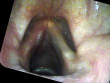
Two weeks after surgery, there is some scar tissue present.
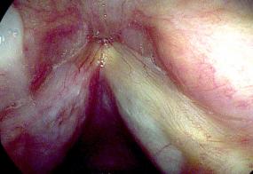
Six months later, there is still no visible regrowth of the lesion.
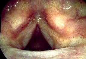
This is an example of vocal cord leukoplakia. That is, a white patch on the vocal cords. This one grew over time. The pathology is benign or not a cancer.

