Cutaneous sarcoid
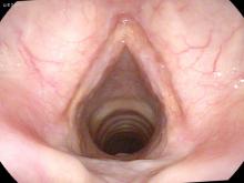
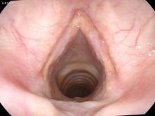
Patient with two years of hoarseness. She has sarcoid of her skin for many years which has been treated with steroid injections.


She underwent biopsies of her vocal cords which showed non-caseating granulomas and chronic inflammation consistent with sarcoid.
Operative photos:
30 endoscope view

70 degree endoscope view

Microscope view

The deposits on her vocal cords keep them from coming together completely during phonation. The deposits are also firm and do not vibrate well during stroboscopy. She also has deposits beneath the vocal cords along the cricoid cartilage.
Several months after the laser treatment of the right vocal cord margin, her voice is improved.


